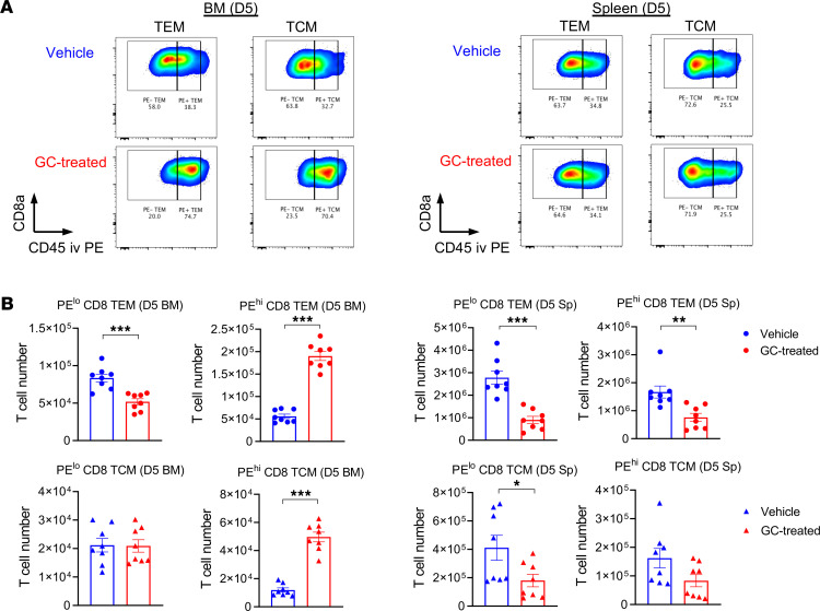
Figure 7. Glucocorticoid treatment promotes donor CD8+ T cell migration to the BM.
(A and B) B6D2F1 mice were transplanted with 2 × 10⁶ purified B6.CD45.1+CD3+ T cells with or without GC treatment. On day 5 after transplantation, recipient mice were injected with a fluorescent-labeled anti-CD45 PE antibody by i.v. injection 5 minutes before euthanasia. (A) Representative FACS plots of CD45 expression on the gated donor CD8+ T cells in the BM (left) and spleen (right) from the vehicle- and GC-treated recipients at day 5 after transplantation. (B) Absolute numbers of CD45.1+ resident (PE low) and circulatory (PE high) CD8+ T cells with TEM and TCM phenotype in the BM and spleen (n = 8 per group, combined from 2 experiments). The fraction of peripheral blood–associated T cells was excluded. *P < 0.05; **P < 0.01; ***P < 0.001. All quantified data are presented as mean ± SEM. Student’s t test with Welch’s modification.

