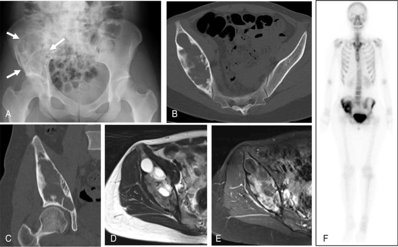
Figure 1.
Images at initial visit (A) Plain radiograph showing a large osteolytic lesion (arrows) on the right ilium. (B) Axial view of pelvic computed tomography (CT) showing an expansile lytic lesion with cortical thinning. (C) Coronal CT showing the lesion extending to the subchondral bone of the acetabulum. (D) Axial T2-weighted magnetic resonance (MR) imaging revealed a multilocular lesion with high signal intensity, and (E) T1-fat suppressed axial MR image showing the lesion to be heterogeneously enhanced with gadolinium. (F) Bone scintigraphy showing increased focal uptake in the right ilium.

