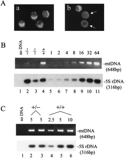
FIG. 4.
Mitochondrial staining and mtDNA copy number analyses. (A) Rhodamine 123 staining of blastocysts. Blastocysts were stained with rhodamine 123 as described in Materials and Methods. Four blastocysts from a wild-type mating (a) are compared to three blastocysts from a heterozygous mating (b). Those with weak fluorescence are indicated by the arrows. (B) mtDNA copy number in blastocysts. Half of the DNA isolated from each of the indicated embryos (lanes 2 to 4) was used to amplify a mtDNA fragment and a 5S rDNA fragment in a PCR. For standards, genomic DNA prepared from the heart of an adult wild-type mouse was diluted and subjected to the same PCR amplification (lanes 5 to 11). The amount of template was indicated in fold at the top for each standard. Products were resolved on a 1.2% agarose gel, and the mtDNA product was visualized by ethidium bromide staining. The 5S rDNA product was detected by Southern blotting. (C) mtDNA copy number in unfertilized eggs. Unfertilized eggs were isolated from wild-type or heterozygous NRF-1 females for DNA preparation as described in Materials and Methods. DNA from 5 eggs from a heterozygous animal (lanes 2 and 3) or from the equivalent of 2.5, 5, or 10 eggs from a wild-type animal (lanes 4, 5, and 6, respectively) was used as the template in PCR amplifications of the same mtDNA sequence as described in panel B. The numbers of eggs from which the template DNA was isolated are indicated above the panels.

