
Figure 1.
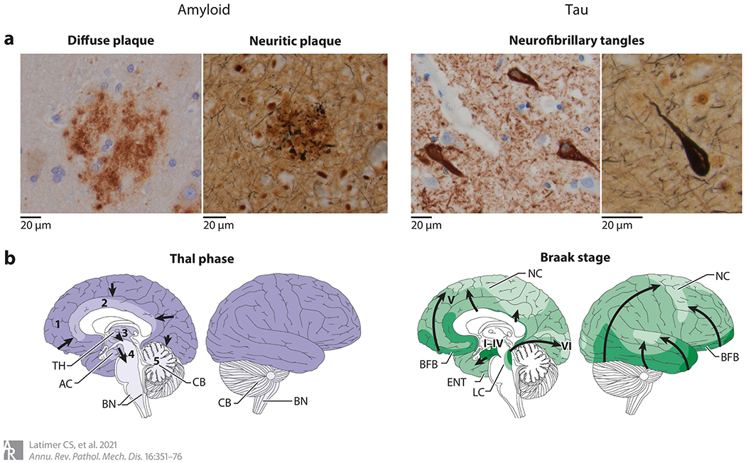
Neuropathologic changes of Alzheimer’s disease. (a) The hallmark features of Alzheimer’s disease neuropathologic change include extracellular amyloid beta (Aβ) plaques and intraneuronal neurofibrillary tangles of hyperphosphorylated tau. Aβ plaques include diffuse forms, highlighted by an anti-Aβ immunostain (left), and neuritic forms, characterized by the inclusion of dystrophic neurites containing hyperphosphorylated tau and highlighted by Bielschowsky silver stain (right). Neurofibrillary tangles can be identified with immunostains to hyperphosphorylated tau (left) as well as silver stains (right). Both amyloid and tau pathology appear to progress throughout the brain in stereotypical patterns. (b) Amyloid progression (purple), as described by Thal phases 1–5 and denoted by arrows, begins in the neocortex (phase 1), followed by limbic regions (phase 2), deep gray nuclei (phase 3), brainstem (phase 4), and, finally, the cerebellum (phase 5). Tau pathology (green), as described by Braak stages I–VI and denoted by arrows, begins in the mesial temporal structures (stages I–IV) before involving neocortex (stage V) and, finally, primary motor and sensory regions (stage VI). Abbreviations: AC, allocortex; BFB, basal forebrain; BN, brainstem nuclei; CB, cerebellum; ENT, entorhinal cortex; LC, locus ceruleus; NC, neocortex; TH, thalamus. Panel b adapted with permission from Reference 42; copyright 2015 Springer Nature.