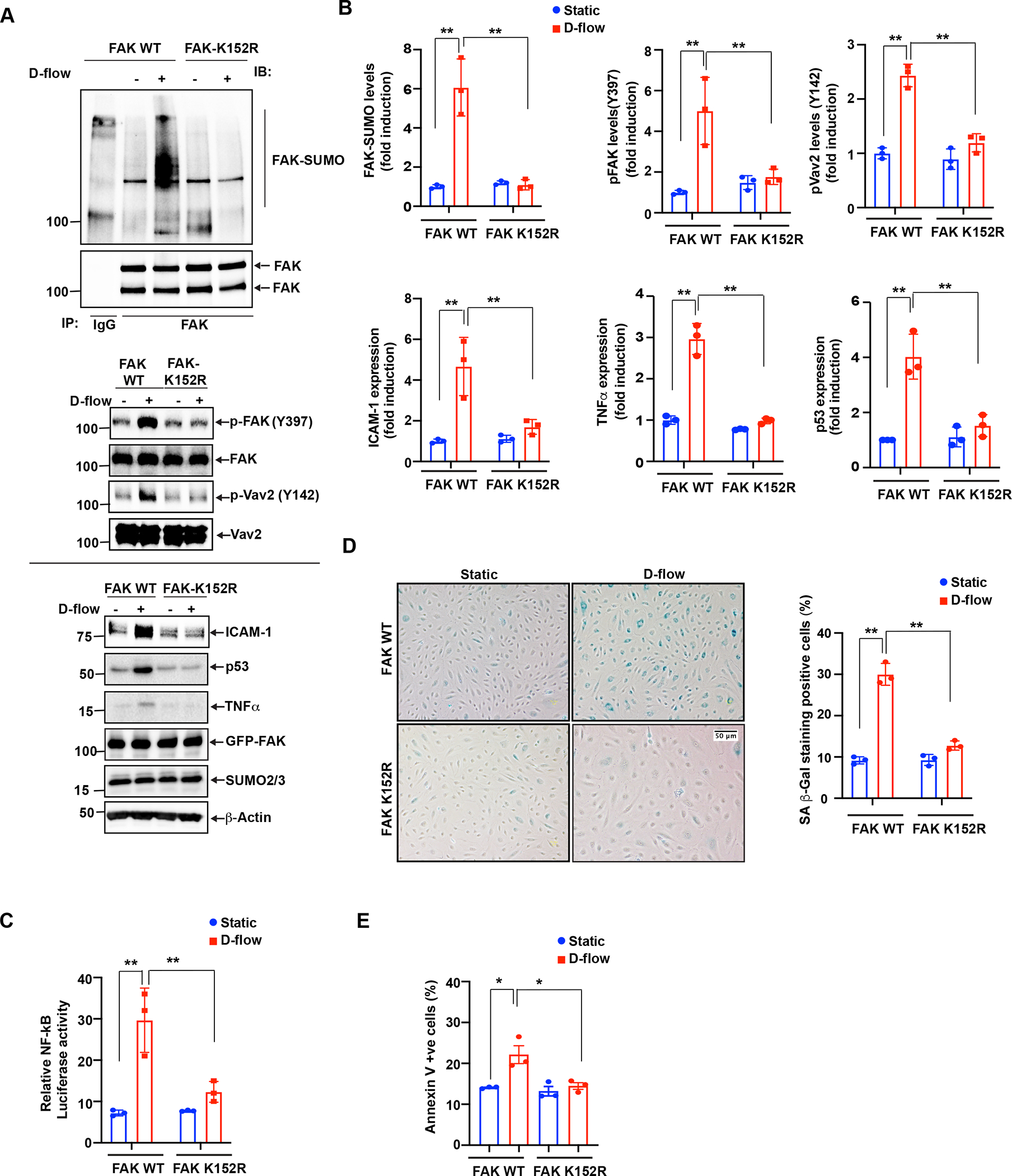
Figure 2: D-flow-induced FAK K152 SUMOylation promotes endothelial cell inflammation and senescence.

(A) HUVECs were transfected with FAK-WT or FAK-K152R plasmids for 36 hours and exposed to D-flow for 30 min or 24 hrs (for ICAM-1, TNF-α, and p53 expression), and cell extracts were prepared. To detect FAK SUMOylation, immunoprecipitation with anti-FAK was performed, followed by immunoblotting with Sumo2/3 antibody. Cell lysates were also immunoblotted for phosphorylation of FAK (p-FAK) and Vav2 (p-Vav2). The blots were sequentially reprobed with anti-FAK, anti-Vav2, anti-Sumo2/3 and β-actin for normalization and loading control. Shown is a representative set of data from three biological replicates. (B) Graphs show densitometric quantification of SUMOylated and phosphorylated FAK, phosphorylated Vav2 and ICAM-1, TNF-α, and p53 expression levels. Fold increases are shown after normalization using the total FAK and Vav2 and β-actin band intensity. Data represent mean ± SD, n = 3, **P < 0.01. (C) HUVECs were transfected with a mixture of either FAK-WT or FAK-K152R plasmid and the dual luciferase NF-kB activity reporter gene and exposed to D-flow for 24 hr. NF-kB activity in cell lysates was measured using the dual-luciferase reporter assay system. Relative NF-κB activity was determined by normalizing firefly luciferase activity to Renilla luciferase activity. Mean±SD (n=3) **P<0.01. (D) HUVECs transfected with FAK-WT or FAK-K152R were exposed to D-flow for 24 hr, and cell apoptosis was determined by annexin V staining as described in Materials and Methods. The graph shows the percent of cells in culture with annexin V staining. Mean±SD (n=3) *P<0.01. (E) HUVECs transfected as described in D were exposed to D-flow for 24 hr. Cells were stained for senescence-associated β-galactosidase (SA-βgal). The graph shows the percentage of SA-B gal positive cells. Mean±SD (n=3) **P<0.01.
