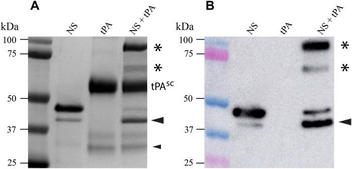
Fig. 1. Detection of covalently linked NS-tPA complexes.
(A) Nonreducing 10% SDS-PAGE analysis of purified wt-NS (NS), tPA, and a mixture of NS and tPA. Molecular markers are indicated on the left side of the image, and the sample loaded into each well is indicated at the top of the image. The position of sc tPA (tPAsc) is indicated; the Actilyse tPA preparation also appears to contain small amounts of twin-chain tPA (tPAtc) dissociated into the A and B chains (lowest two bands migrating at ~31 and 28 kDa, respectively). When NS was incubated with tPA, two high–molecular weight, SDS-stable species formed (asterisks) and an increase in cleaved NS was also apparent (large black arrowhead). The small black arrowhead indicates the position of the B chain of tPAtc containing the serine protease domain that reacted with NS to produce the faster-migrating NS-tPA complex (lower asterisk). (B) Immunoblotting using an antibody specific for NS confirmed that the high–molecular weight complexes (asterisks) contained NS and only form when NS is incubated with tPA. The larger complex (upper asterisk) corresponds to one containing tPAsc, while the smaller complex (lower asterisk) corresponds to one formed between NS and the B chain of tPAtc.

