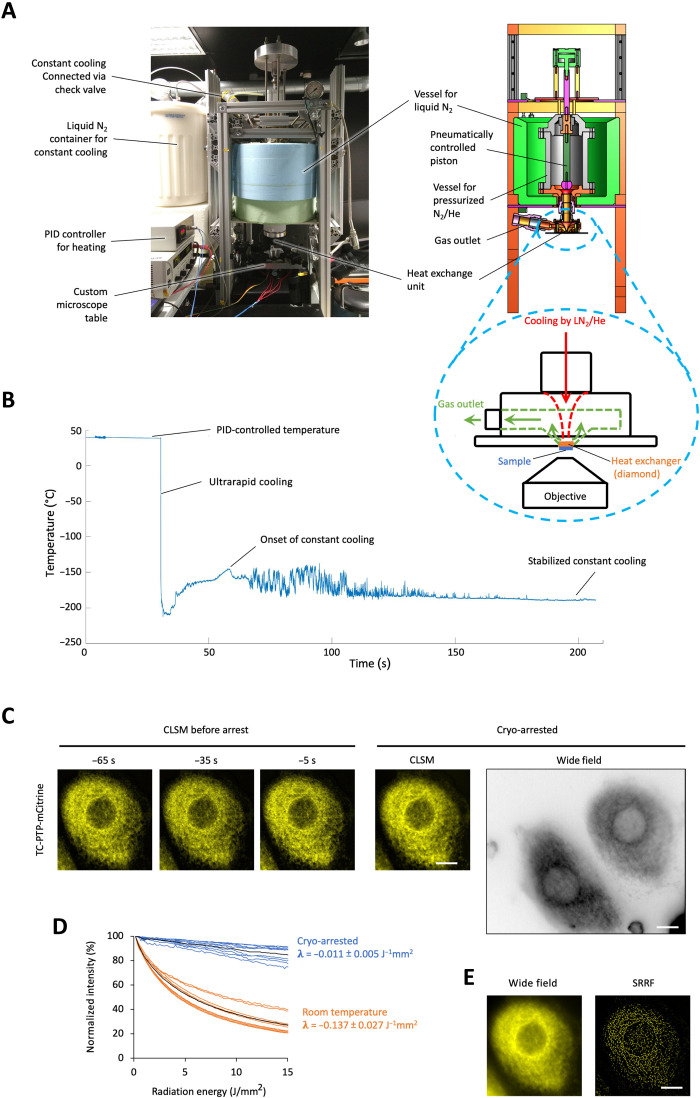
Fig. 1. Ultrarapid cryo-arrest microscopy.
(A) Photograph and schematic of ultrarapid cooling device. Cyan dashed circle: heat exchanger unit; red arrow: flow of liquid nitrogen (LN2) with gaseous He toward diamond heat exchanger; green arrows: expanded gas outflow. The whole cooling device is lowered above an epifluorescence microscope objective. (B) Measured temperature course (50-μm constantan-copper thermocouple in 100-μm aqueous sample). (C) CLSM of TC-PTP-mCitrine in a MCF7 cell before (at indicated time, 3.7-s scan time) and during cryo-arrest (10 frames, 37-s scan time). Right: Corresponding wide-field fluorescence image shown in inverted gray scale to visualize dim extensions of the endoplasmic reticulum. (D) mCitrine photobleaching at room temperature (orange; N = 9) and under cryo-arrest (blue; N = 9); 100 wide-field frames. λ: photobleaching rates (mean ± SD). (E) Sum (left) and SRRF reconstruction (right) from 100 wide-field frames under cryo-arrest. Scale bars, 10 μm.

