Figure 1.
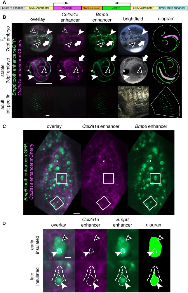
An insulated bicistronic construct reports separate expression patterns from two different enhancers. (A) Bicistronic construct with a Col2a1a enhancer and Hsp70l promoter driving mCh and the freshwater Bmp6 intronic tooth enhancer and Hsp70l promoter driving eGFP, separated by the mouse tyrosinase insulator (GAB). (B) Transgenic fish show a separation of domains in red and green overlay, red channel only, green channel only, brightfield, and diagram (left to right). Top: In 7 days post fertilization (dpf) F0 embryos (dorsal view), insulation was observed in some but not all domains. Both mCh and eGFP were observed in the same area in the right pectoral fin (dotted arrowhead), indicating incomplete or failed separation of domains, while in other areas of the pectoral fin only eGFP was observed (black arrowhead). Within the notochord (solid white arrowhead), only mCh was observed, while in the median fin (white arrow) only eGFP was observed, indicating insulation in both domains. Middle: In 7 dpf stable F1 embryos (lateral view), only eGFP was observed in the pectoral fins (black arrowhead) indicating successful insulation in those domains, while both fluorophores were detected in the median fin (white arrow) and in the notochord (solid white arrowhead) indicating a lack of insulation. Both fluorophores were detected in the lens of the eye (asterisk), a domain driven by the Hsp70l promoter. Bottom: in adult pectoral fins (lateral view), eGFP but not mCh expression was detected. Diagram: schematic of eGFP and mCh expression in fins and notochord, with overlap shown in gray. Spheres trace the outline of the chorion (top, middle), and white lines trace the pectoral fin (bottom). (C and D) Dorsal pharyngeal tooth plate (C) and representative teeth of early and late stages (D) from adult stable transgenic fish. (C) Insulator effectiveness was observed with eGFP restricted to predicted tooth domains and mCh primarily present in the surrounding tissue. In some teeth, faint mCh appeared to be expressed in the dental mesenchyme (asterisk). (D) eGFP expression was detected in the dental mesenchyme (solid arrowhead and extent of mesenchyme as white dotted line) and dental epithelium (black arrowhead) of developing teeth, while mCh was expressed in the surrounding tissue (white dashed line outlines a mineralized tooth). Scale bars=1 mm (B), 100 µm (C), 25 µm (D). n=92 F0 embryos, >50 F1 embryos, >3 adult fish per time point and 3 teeth per fish for adult stage.

