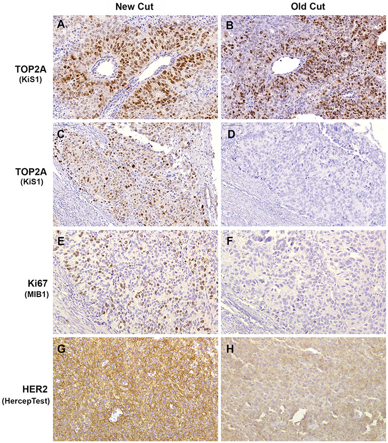
Fig. 2.
Photomicrographs of comparative TOP2A, Ki67 and HER2 IHC staining of new cut (3 weeks old) and stored (old cut, >10 years old) slides sectioned from the same FFPE tumor blocks of endometrial cancers. (A & B) Example of endometrial cancer demonstrating no effect of slide storage on TOP2A IHC interpretation. Strong (3+) positive nuclear staining was observed in both sections. (C – F) Staining patterns of TOP2A (C & D) and Ki67 (E & F) illustrating complete loss of immunoreactivity in old cut slides of the same case. In new cut sections of this tumor, the percentages of positively stained cells were 50% for TOP2A and 30% for Ki67. (G & H) Tumor showing change in HER2 intensity staining from strongly positive (3+; G) to weak (1+; H). Stroma and inflammatory cells were negative internal controls for all biomarkers. Original magnification x200.

