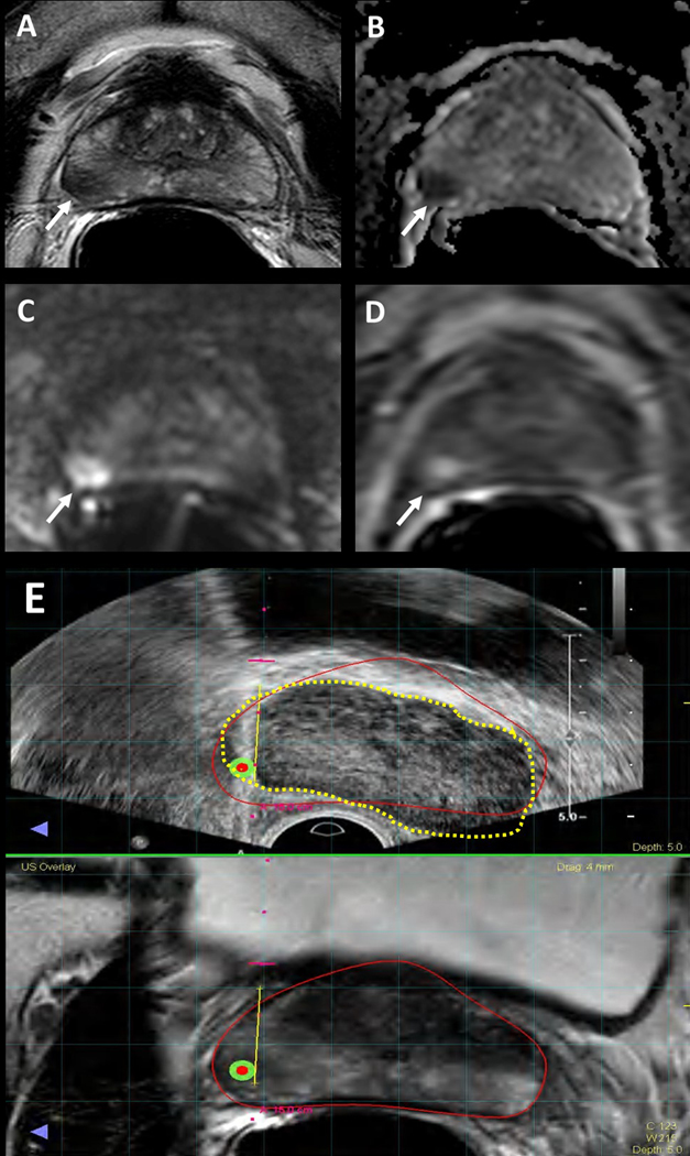
Figure 1.
Multiparametric MRI image example of clinically significant cancer that was missed due to MRI-targeted biopsy targeting error. 56-year-old male with a serum PSA of 5.9ng/ml. Axial T2-Weighted (T2W) MRI shows a hypointense lesion in the right mid-peripheral zone (arrow) (A), the lesion shows diffusion restriction on ADC map (B) and b2000 DW MRI (C) and early enhancement on DCE MRI (D) (arrows). Targeted biopsy revealed Gleason 3+4 prostate cancer in this lesion, whereas systematic biopsy yielded Gleason 4+4 prostate adenocarcinoma in the right peripheral zone. (E) Screen captures of the TRUS/MRI fusion guided biopsy procedure (E) demonstrates a registration error between MRI (inked in red) vs. TRUS (inked in dashed yellow), which is likely the reason for the under-sampling of the right mid peripheral zone lesion during TRUS/MRI fusion guided biopsy.

