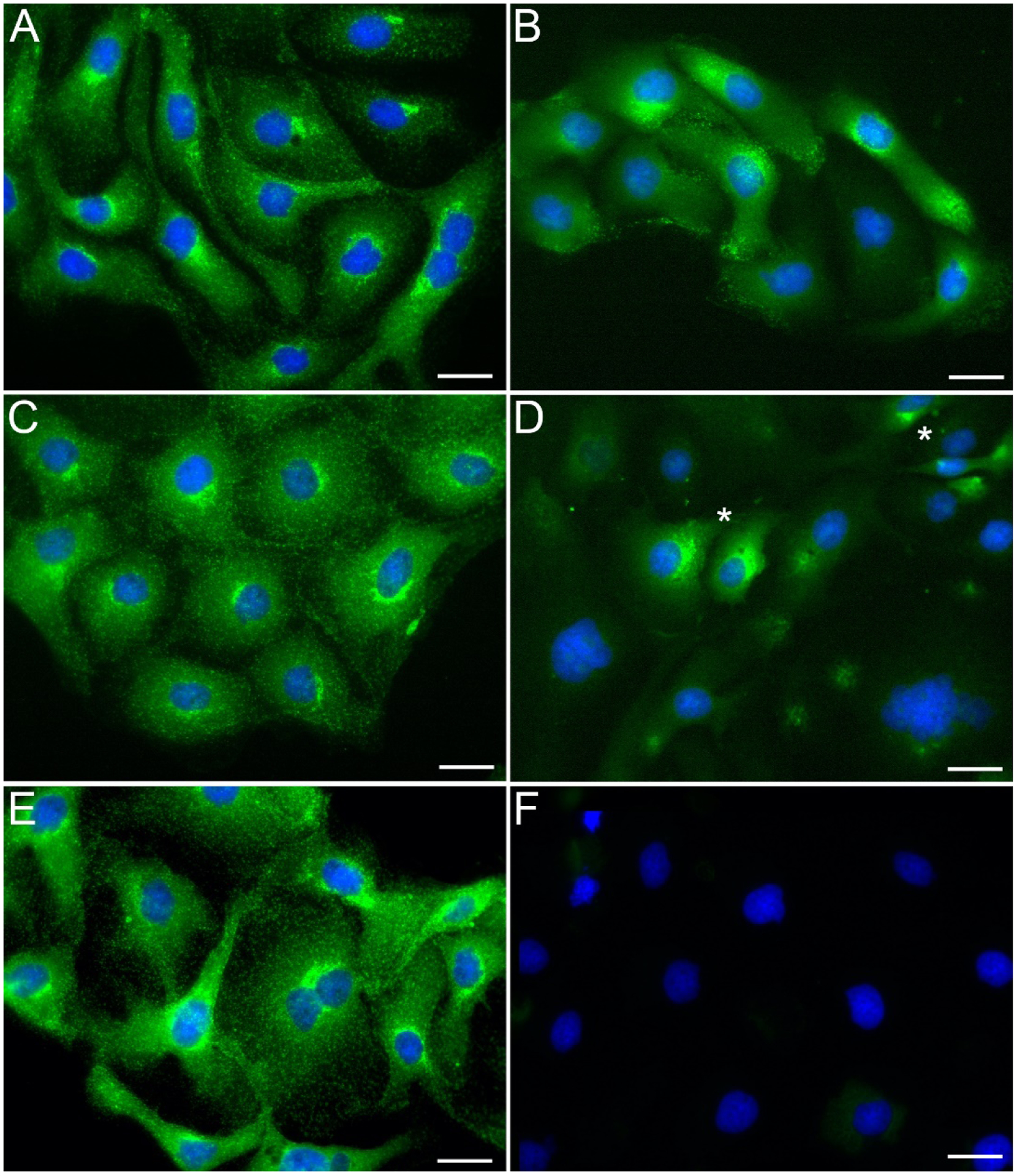
Figure 1. The buffer formulation affects the plasma membrane permeability.

Following our RCIA protocol, we fixed PTK-1 cells in a 4% solution of paraformaldehyde prepared in PBS (A, B), DPBS (C, D) or PHEM buffer (E, F; see Methods for formulation) and then immunostained them in the respective buffer after treatment with detergent (0.1% Triton X-100, 5min before adding primary antibody; A, C, E) or without detergent added (B, D, F) using a mouse monoclonal antibody against the cytosolic epitope of clathrin, followed by a goat anti mouse, Alexa 488 (displayed in green). Cells were counterstained with DAPI after completion of the immunostaining (displayed in blue). All three buffers supported specific binding of the primary antibody when used in conjunction to Triton X-100 (A, C, E). When the detergent was omitted, specific binding to the antigen was observed in most cells in presence of PBS (B), and to a lesser extent in presence of DPBS, along with occasional polarized localization in few cells (D; asterisks); in presence of PHEM however, the staining was non-detectable (F). Scale bars, 10μm.
