
Fig. 5.
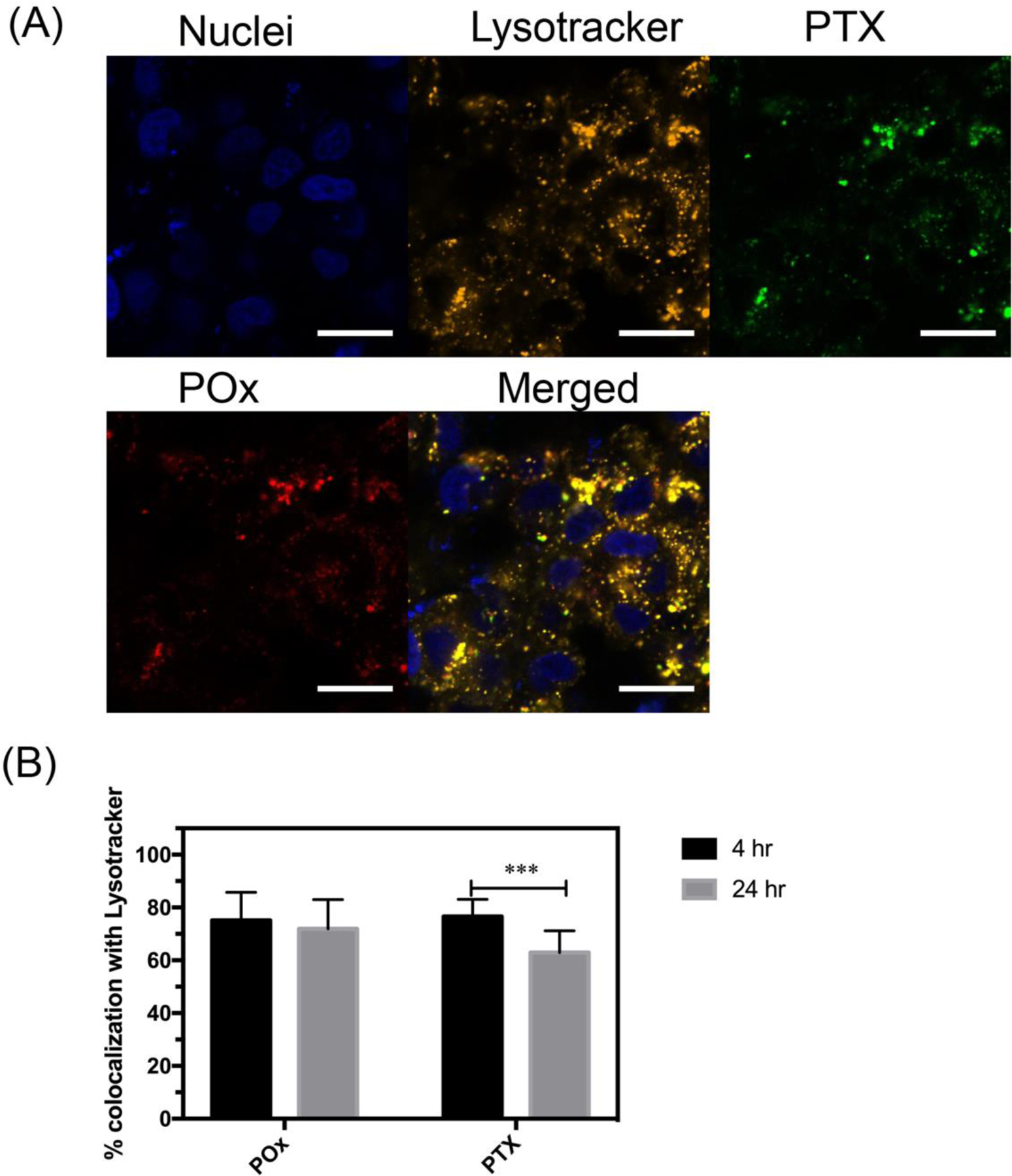
(A) Intracellular distribution of fluorescently labeled PTX-NFG/B in MCF-7 breast cancer cells after 4 h of incubation (bar = 20 μm). (B) Quantification of the colocalization of AF647-POx (red) and OG488-PTX (green) with lysosomes (orange) as determined by ImageJ software after 4 and 24 h of incubation (***P<0.001). Nuclei were stained with Hoechst 33342 (blue).