
Figure 5. Colocalization of activated β-catenin and YAP is reduced in the presence of Wnt inhibitors on stiffer MC-GAG materials.
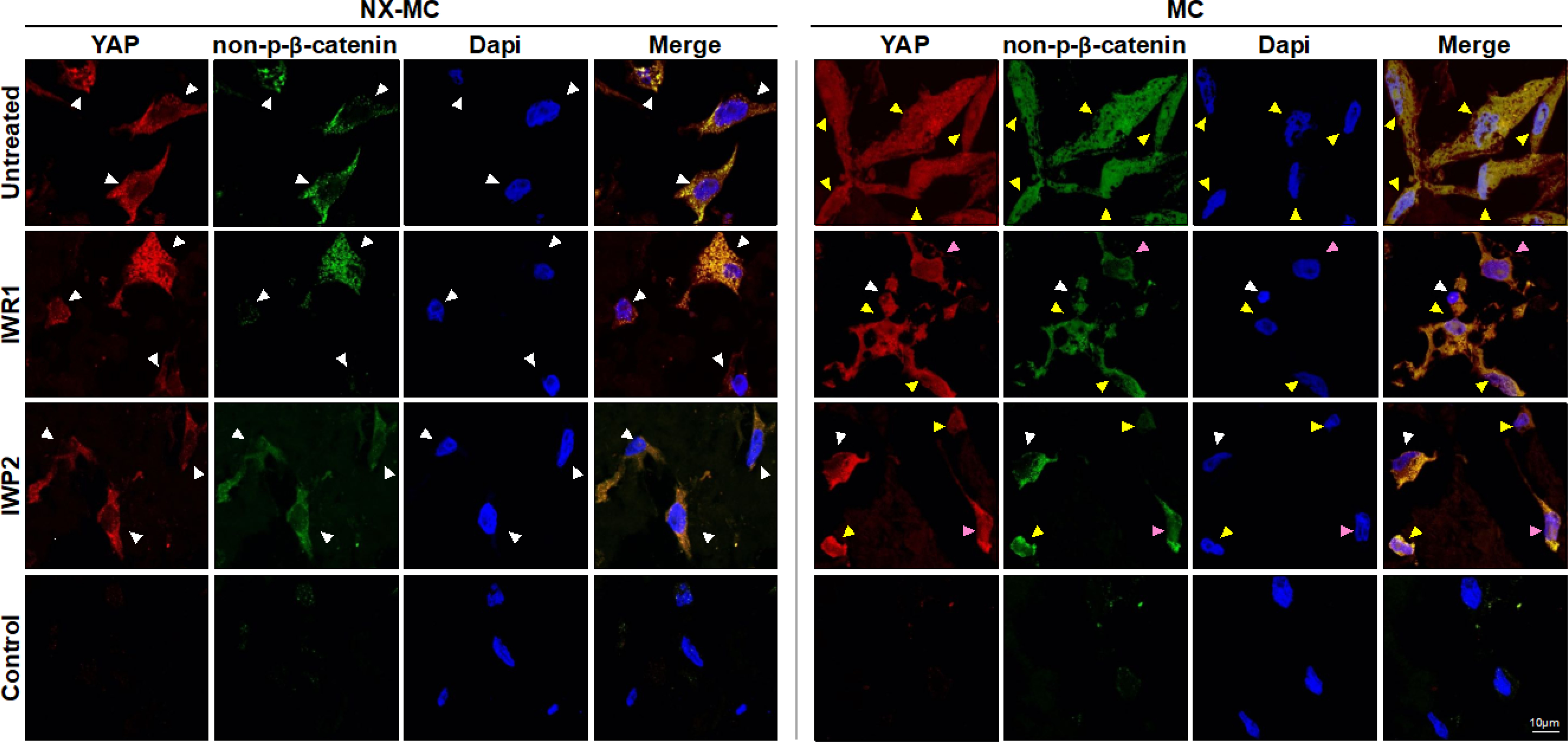
Representative confocal microscopic images of primary hMSCs cultured on NX-MC (left panels) or MC (right panels) for 7 days untreated (top row) or treated with 50 μM IWR1 (second row) or IWP2 (third row) and stained for YAP or non-p-β-catenin. Negative control (Control) using secondary antibody only and Dapi on cells cultured on MC shown on bottom row. Scale bar indicates 10 μm. White arrows indicate cells with primarily cytosolic staining, yellow arrows indicate cells with both cytosolic and nuclear staining, and pink arrows indicate cells where YAP and non-p-β-catenin demonstrate a reduction in colocalization.