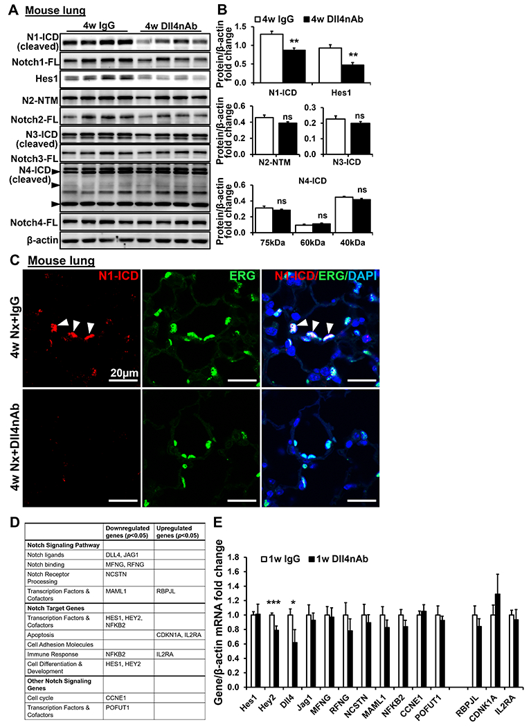
Figure 2. Decrease of N1-ICD expression by 4w Dll4nAb treatment in mice lung tissues and inhibition of Notch1 cleavage induces Notch1 target genes dysregulation in ECs.

(A-B) N1-ICD, Notch1, Hes1, N2-NTM, Notch2, N3-ICD, Notch3, N4-ICD, and Notch4 were detected in lungs of Dll4nAb and control IgG injected mice by western blot (A) and quantified (B) (n=15). Proteins normalized to β-actin. (C) Double immunofluorescence staining of N1-ICD (red) and ERG (green) on lungs of Dll4nAb and control IgG injected mice(n=5). Arrows indicate N1-ICD staining. Scale bars: 20μm. Nuclei were counterstained by DAPI (blue). (D) EC specific Notch target genes were measured by PCR Arrays(n=3). (E) EC specific Notch target genes were measured in lungs of 1w Dll4nAb injected mice and control mice (n=6) by qPCR. Data shown as Mean±SEM; P values were calculated using the Student’s t-test, and *P<0.05; **, P<0.01; ***, P<0.001 vs control IgG group. ns, not significant.
