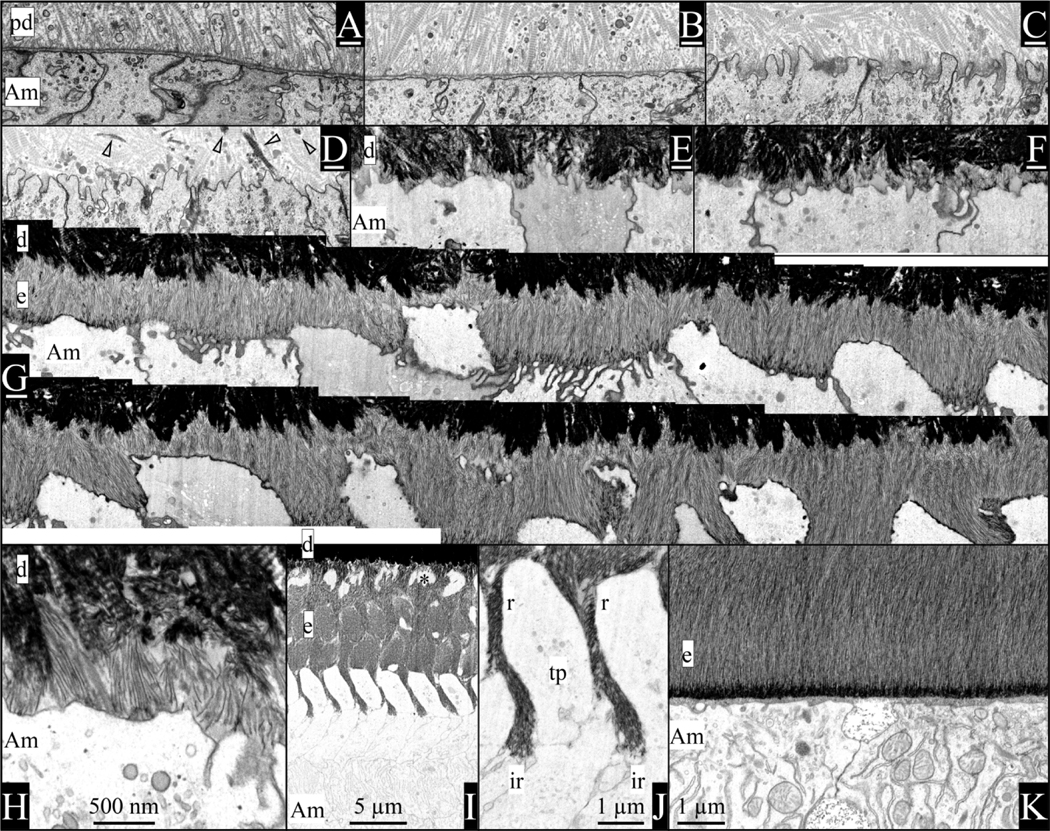
Fig. 1.
Visualization of enamel formation by focused ion beam-backscattered scanning electron microscopy (FIB-bSEM) of a continuously growing 7-week mouse mandibular incisor. A) Ameloblasts are separated from the unmineralized predentin matrix by a well-defined basement membrane (BM). The predentin matrix contains odontoblastic processes, matrix vesicles released by odontoblasts, and numerous collagen fibrils/fibers that are oriented toward the ameloblasts where they appear to attach to the BM. B) The BM thins as the ameloblast membrane becomes less linear. C) Ameloblast finger-like processes penetrate the BM and extend into predentin between the collagen fibrils, as remnants of the BM accumulate along the ameloblast membrane that is reabsorbing it. D) Mineral nuclei appear in predentin some distance from the ameloblast membrane (arrowheads). E) Dentin mineral coalesces into a continuous mineral layer that extends to near the ameloblast membrane. F) Enamel ribbons initiate in patches on dentin mineral associated with the ends of mineralized collagen fibers. G) A continuous segment of mouse incisor completing the formation of initial enamel (above the G) and showing the progressive formation of Tomes processes as the segment continues in a second row below the G. The ends of Tomes’ processes form by the rapid extension of “prongs” of mineral ribbons at interrod growth sites along the peripheral part of the distal ameloblast membrane near the cell junctions. H) High magnification of initial enamel ribbons forming on the ends of collagen fibers and extending at different angles back to the ameloblast membrane, following the finger-like process as it retreated into the cell membrane. I) Rod and interrod enamel after formation of the Tomes process [asterisk indicates a space of Weber (Bartlett et al., 2021)]. J) Tomes’ process showing the positions of rod (r) and interrod (ir) growth sites in the mineralization front. K) Maturation stage enamel has a basal lamina comprised of SCPP proteins bound to the surface enamel. Key: Am, ameloblast; d, dentin, e, enamel, ir, interrod growth site; pd, predentin; r, rod growth site; tp, Tomes’ process; white scale bars (under figure letters) = 500 nm.

