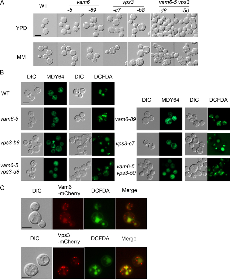
Figure 1. Vps3 and Vam6 are localized on endosomes and influence vacuolar and cell morphology.
(A) Differential interference microscopy (DIC) to examine the cell morphology of the WT, vps3Δ, vam6Δ and vps3Δ vam6Δ deletion strains. Cells were harvested from the overnight cultures in either yeast extract peptone dextrose (YPD) or minimum medium (MM). The absence of either VPS3 or VAM6 resulted in a similar morphology to the WT strain. Cells lacking both VPS3 and VAM6 displayed swollen or ecliptic shape and tended to be unseparated. (B) Vacuolar morphology in the WT strain and the indicated mutants. Vacuoles were stained with MDY64 or 5-(and-6)-carboxy-2’,7-dicholorofluorescein diacetate (carboxy-DCFDA). (C) Localization of Vam6-mCherry and Vps3-mCherry in WT cells upon growth of cells in minimum medium (MM). Vacuoles were stained with carboxy-DCFDA. Bar = 5 μm in all images.

