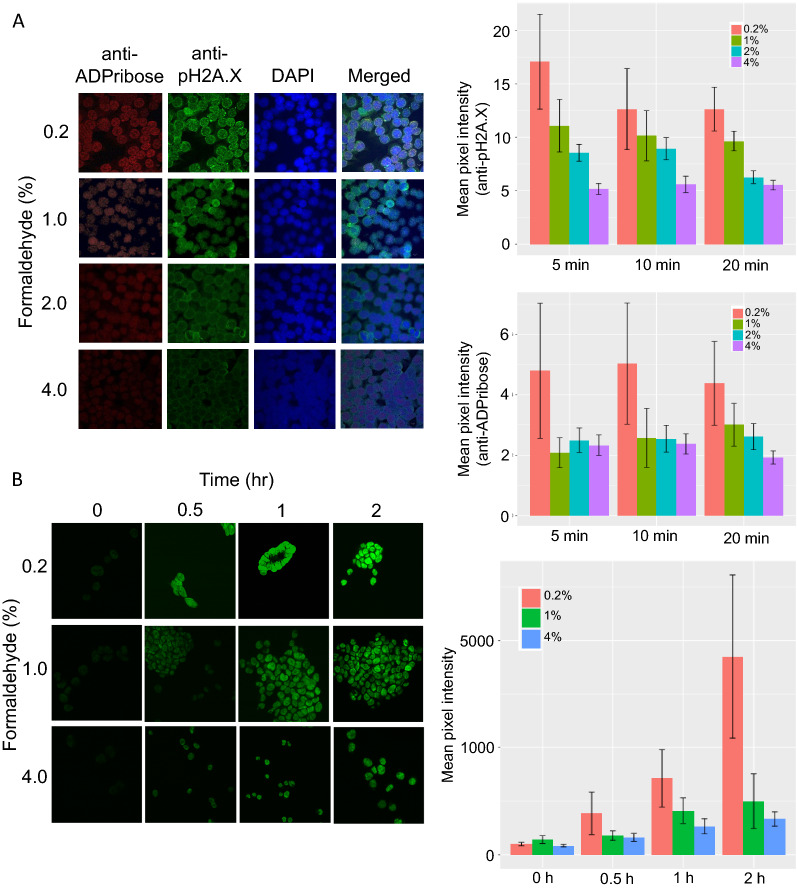
Fig. 1.
Formaldehyde fixation condition, DNA damage response pathways markers and one-pot UniNicE-seq labeling efficiency in HCT116 cells. A Left panel, representative visualization of ADPribose and phosphoSer139 histone H2A.X using 561 nm (red) and 488 nm (green) laser of cells fixed with different concentration of formaldehyde as indicated in Y axis. The antibodies used for probing are indicated on top. DNA content of nuclei is revealed by DAPI (blue) staining. Right panel shows quantification of fluorescence, represented as mean pixel intensity from anti-pH2A.X and anti-ADPribose staining. B Visualization of NicE-view using fluorescein-dATP (green) on cells differently fixed with different formaldehyde concentrations (Y-axis) at different time intervals indicated on the top. Left panel shows Fluorescein-12-dATP labeling in a one-pot UniNicE-seq reaction. The labeling efficiency is shown at right panel

