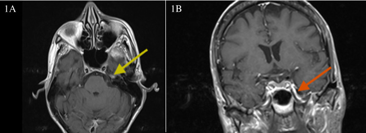
Figure 1. T1 post-contrast weighted MRI images of the brain.
1A: Axial T1 post-contrast weighted MRI images with gadolinium enhancement of the semilunate Gasserian ganglion (yellow arrow). 1B: Coronal T1 post-contrast weighted MRI images demonstrating mandibular nerve enhancement in the foramen ovale (red arrow)
MRI: magnetic resonance imaging

