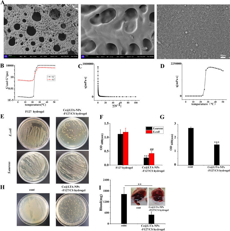
Fig. 8.
Morphology, rheology and antibacterial properties of Ce@LTA-NPs-F127/CS hydrogels. A Representative SEM image of the Ce@LTA-NPs-F127/CS hydrogels. B Storage and loss modulus of Ce@LTA-NPs-F127/CS hydrogels with change of temperature. C The viscosity of Ce@LTA-NPs-F127/CS hydrogels with the changes of shear stress. D The viscosity of Ce@LTA-NPs-F127/CS hydrogels with the changes of temperature. E Photos of E. coli and S. aureus culture plates after exposure to Ce@LTA-NPs-F127/CS hydrogels. F OD600nm of bacterial suspension treated with Ce@LTA-NPs-F127/CS hydrogels. These data represent three separate experiments and are presented as the mean values ± SD. ##p < 0.01, ***p < 0.001. G OD600nm of bacteria in wound exudates from diabetic wound tissues treated with Ce@LTA-NPs-F127/CS hydrogels. OD600 nm of bacteria in wound exudates from untreated diabetic wound tissues was set as control group. These data represent three separate experiments and are presented as the mean values ± SD, ***p < 0.001. H Photos of bacteria in wound exudates from diabetic wound tissues treated with Ce@LTA-NPs-F127/CS hydrogels. Photo of bacteria in wound exudates from untreated diabetic wound tissues was set as control group. I Haemorrhagic analysis in liver treated with Ce@LTA-NPs-F127/CS hydrogels. The amount of bleeding after applying the hydrogels in the bleeding site of liver and comparing the results with the untreated bleeding site (control group). These data represent three separate experiments and are presented as the mean values ± SD, **p < 0.01

