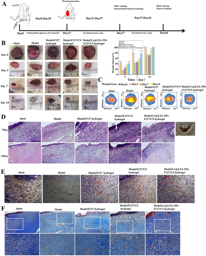
Fig. 9.
Healing process and histological evaluation of diabetic wound promoted by Ce@LTA-NPs-F127/CS hydrogels dressing. The untreated wound in the normal rat was set as sham group. The untreated wound in a type 1 diabetic rat was set as model group. A Design chart of animal experiment. B Representative images and wound healing rate of full-thickness skin defect in a type 1 diabetic rat model treated with F127 hydrogels, F127/CS hydrogels, Ce@LTA-NPs-F127/CS hydrogels after operation. These data are represented as the means ± SD (n = 3). *p < 0.05, **p < 0.01, ***p < 0.001. C Traces of the wound closure during 14 days for each treatment. D Representative H&E staining images of wound sections treated with F127 hydrogels, F127/CS hydrogels, Ce@LTA-NPs-F127/CS hydrogels at 7th day and 14th day after operation. The scale bar is 100 μm. E Immunohistochemistry assay of CD31 expression in diabetic wound sites treated with F127 hydrogels, F127/CS hydrogels, Ce@LTA-NPs-F127/CS hydrogels at 7th day. Vascular endothelial cell (CD31) and cell nucleus were stained brown and blue. Neovascularization was identified by positive CD31 staining (brown). The scale bar is 50 μm. F Masson’s trichrome staining of wound sections treated with F127 hydrogels, F127/CS hydrogels, Ce@LTA-NPs-F127/CS hydrogels at 14th day after operation. The scale bar is 100 μm

