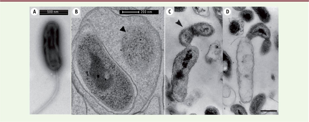
Fig. 1. Transmission electron microscopic observation of endobiotic predation by B. bacteriovorus H100 (B) and epibiotic predation by B. exovorus (C & D.
B. bacteriovorus is shown within the periplasm of an E. coli cell (B) and B. exovorus attached externally to the organism (C & D). One can observe the reduced cytoplasm of the prey organism (B arrow), the extracellular growth and division of the epibiotic predator (C arrow), and the emptied body of the prey organism (D). Reproduced from [3] with permission

