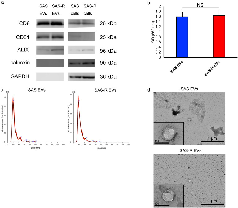
FIGURE 1.

Characterization of OSCC cells derived EVs. (a) Western blotting of EVs and whole‐cell proteins was performed to confirm exosome marker proteins (CD9, CD81, ALIX), calnexin, and GAPDH. (b) The colorimetric BCA protein assay was performed to measure the EV protein. (c) The size distribution and the concentration of the EVs were measured with Nanoparticle Tracking Analysis. (d) Transmission electron micrograph (wide‐field and close‐up) of SAS EVs and SAS‐R EVs. Values are expressed as mean ± standard deviation of triplicate samples. EV, extracellular vesicles; OSCC, oral squamous cell carcinoma; GAPDH, glyceraldehyde‐3‐phosphate dehydrogenase; NS, no significant differences
