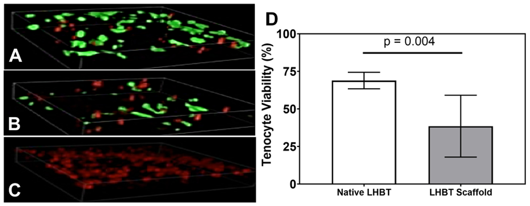
Figure 3. Scaffold formation resulted in decreased viability of resident long head biceps tendon tenocytes.

Representative 3-D confocal z-stack LIVE/DEAD images (400x total mag.) of A) native, B) meshed, and C) ethanol treated (negative control) LHBT samples depicting viable (green) and dead (red) tenocytes within two hours of harvest or expansion. D) Quantitative analysis of LHBT tenocyte viability from LIVE/DEAD images illustrating a significant reduction in resident LHBT tenocyte viability within two hours after LHBT scaffold formation. Horizontal lines indicate a statistical difference (p<0.05) compared to native LHBT. Graphed data represent means and 95% confidence intervals.
