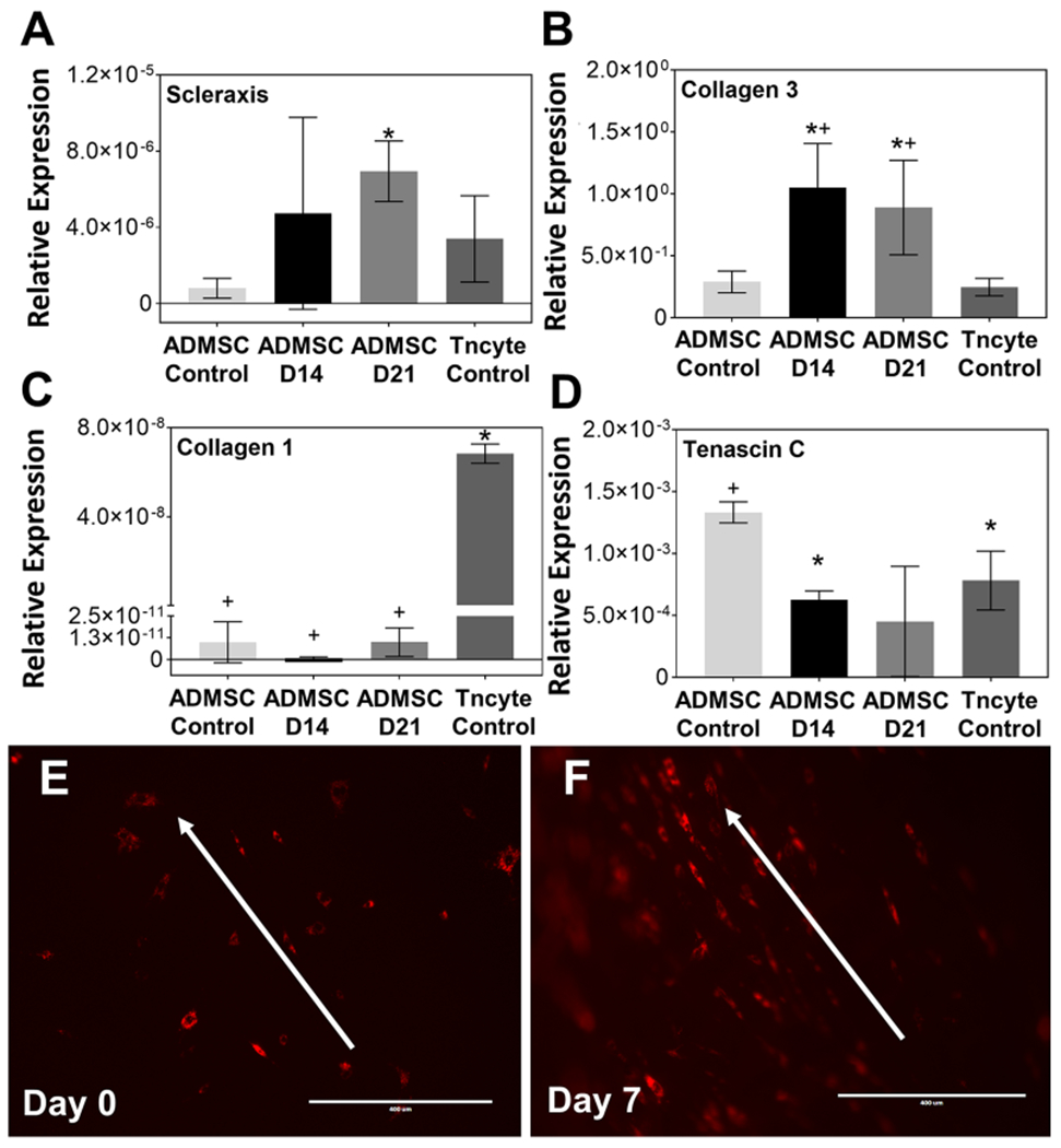
Figure 4. Soluble factors produced by tenocytes derived from long head bicep tendon scaffolds promote tenogenic differentiation of ADMSCs and scaffolds themselves support ADMSC alignment and a tenocyte-like morphology.

Gene transcript (mRNA) expression of A) scleraxis, B) collagen type 3, C) collagen type 1 and D) tenascin c relative to GAPDH for ADMSCs cultured in media conditioned by tenocytes from LHBT scaffolds for 14 (“ADMSC D14) and 21 (“AMSC D21”) days illustrating up-regulation of tenogenic markers Scleraxis and Collagen Type III compared to undifferentiated ADMSC negative controls (“ADMSC Control”). + and * indicate statistical differences (p<0.05) compared to LHBT tenocyte positive controls (“Tncyte Control”) and undifferentiated ADMSC negative controls, respectively. Graphed data represent means and 95% confidence intervals. Fluorescent imaging of ADMSCs cultured on LHBT scaffolds at E) Day 0 (i.e. 2 hours post-seeding) and F) Day 7 illustrating progressive cell alignment and development of spindle-shape morphology along the direction of collagen fibrils (white arrows) in scaffolds. Scale bar = 400μm.
