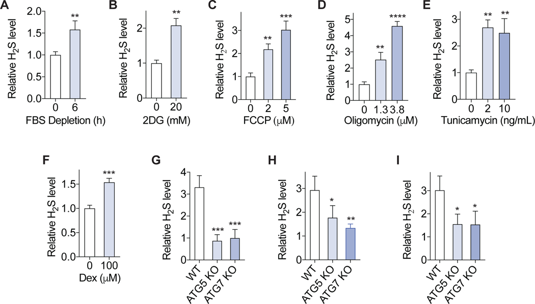
Figure 4. Autophagy-dependent H2S generation is a general response to stress.
(A-F) Primary MEF cells were treated with serum (FBS) depletion (A), 2DG (B), FCCP (C), oligomycin (D), tunicamycin(E), dexamethasone (Dex) (F) for 6h, and intracellular H2S level was measured (n=3). (G-I) Immortalized WT, ATG5 knockout and ATG7 knockout MEF cells were treated with serum depletion (G), 20 μM FCCP (H), 150 μM Dex (I) for 6 h, and intracellular H2S level was measured. The relative H2S levels to the WT cells vehicle control are shown (n=3). All intracellular H2S measurements were performed using probe SF7-AM. Error bars indicate SD. *P < 0.05; **P < 0.01; *** P < 0.001; **** P < 0.0001. Student’s t-test (A, B, F); One way ANOVA (C-E, G-I).

