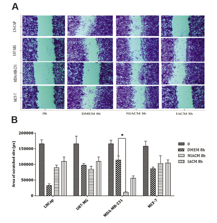
Fig.3.
Wound healing assay at the beginning (0 hour) and 8 hours of different tumor cell lines in different culture media. A. A scratch was made in a confluent monolayer of LN-CAP, U87MG, MDA-MB-231, and MCF-7 cancer cell lines, and they were then cultured in the presence of DMEM, NIACM of ASCs and IACM of ASCs. Images were acquired at 0 and 8 hours post-incubation (scale bar: 100 μm). B. Analysis of wound healing response of cancer cell lines following exposure to DMEM, NIACM, and IACM of ASCs. Data are presented as mean ± SEM. P<0.05 was considered significant. Kruskal-Wallis test was used for the analysis of non-parametric data. ASCs; Adipose-derive mesenchymal stem cells, IACM; Irradiated ASCs-conditioned media, NIACM; Non-irradiated ASCs-conditioned media, h; Hours, DMEM; Dulbecco’s Modified Eagle’s Medium, and *; P<0.05.

