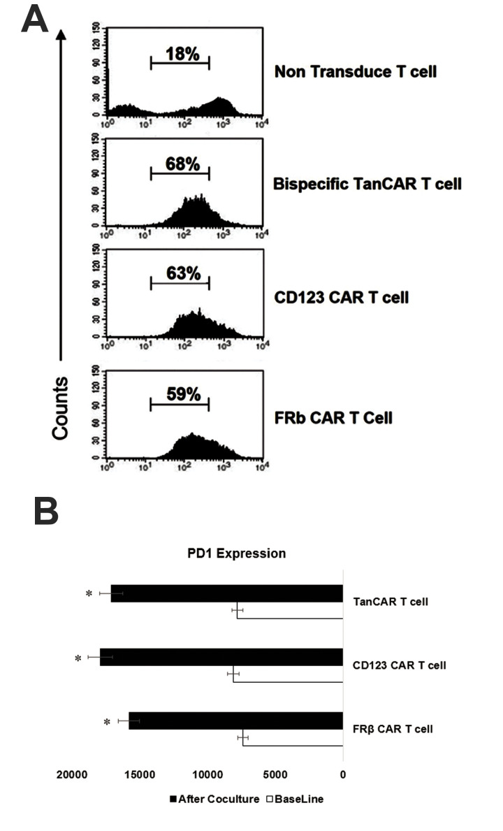
Fig.5.
Assessment of TanCAR-T cell proliferation and expressions of their exhaustion markers PD-1. A. Flow cytometry output during incubation of TanCAR CD123-FRβ, monospecific chimeric antigen receptor (CAR-T) cells, and non-transduced cells with the THP1 leukaemia cell line after 10 days of co-culture. B. Median fluorescence intensity results for expression of PD-1 on the CD8+ CAR-T cells before and after repeated stimulation with the leukaemia cell line for one week. Representative data are from three independent experiments, each done in triplicate. A single-step Tukey’s range test was used. *; P≤0.05.

