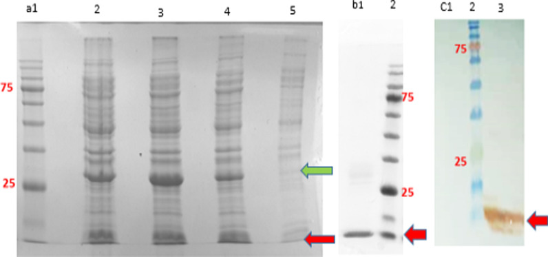
Fig. 1.
The expression analysis of eGFP and SAK proteins in dicistronic SILEX system. a 15% SDS-PAGE analysis of eGFP and SAK expression [Lane 1: protein marker, Lanes 2 and 3: 16 h after inoculation in 1-L culture at two different clones, lane 4: 16 h after inoculation in 5-ml culture, and Lane 5: 2 h after inoculation at 5-ml culture], b 15% SDS-PAGE analysis of purified SAK [Lane1: purified SAK, Lane 2: protein marker], c western blot analysis of SAK protein [Lane 1: E. coli BL21 (DE3) containing pET28a-sak-rbs-egfp (negative control), Lane 2: protein marker, Lane 3: double transformed E. coli BL21 (DE3). The green and red arrows indicate eGFP and SAK proteins, respectively. The protein marker molecular weights are 180, 135, 100, 75, 63, 48, 35, 25, 17, and 11 kDa

