
Figure 6. TRIM5α Interaction with the Flavivirus Protease Is Associated with Virus Restriction.
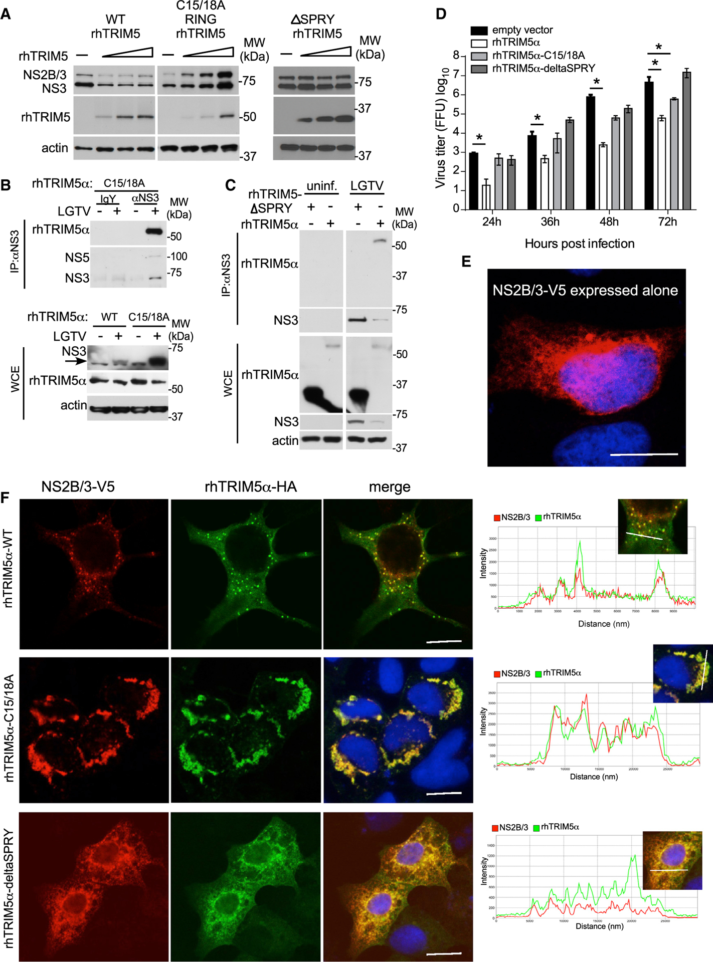
(A) Western blot analysis following transfection of constant amounts of LGTV NS2B/3-V5 plasmid with increasing amounts of rhTRIM5α-HA, RING mutant rhTRIM5(C15/C18A)-HA, or rhTRIM5-delta SPRY-HA, as indicated in HEK293 cells.
(B and C) IP of NS3 from LGTV-infected HEK293 cells (MOI of 0.01; 48 hpi) stably expressing (B) RING rhTRIM5(C15/C18A)-HA or (C) rhTRIM5α-HA or rhTRIM5-delta SPRY-HA.
(D) LGTV replication kinetics in HEK293 cells stably expressing rhTRIM5α-HA, RING mutant rhTRIM5(C15/C18A)-HA, rhTRIM5-delta SPRY, or the empty vector control following infection at MOI of 0.001. All data are from 3 independent experiments in triplicates (mean ± SD, *p < 0.05 Mann-Whitney test).
(E and F) HEK293 cells were co-transfected with LGTV NS2B/3-V5 (shown expressed alone in E), WT rhTRIM5α-HA, RING mutant rhTRIM5(C15/C18A)-HA, or rhTRIM5-delta SPRY-HA (F). Slides were fixed and processed for indirect immunofluorescence staining with antibodies specific for HA (green) and V5 (red), and nuclei were counterstained with DAPI (blue). Images were analyzed using confocal microscopy with fluorescence intensity profiles measured across the white line of insets to demonstrate colocalization using Zen Imaging software. Scale bar, 10 μm. See also Figure S5.