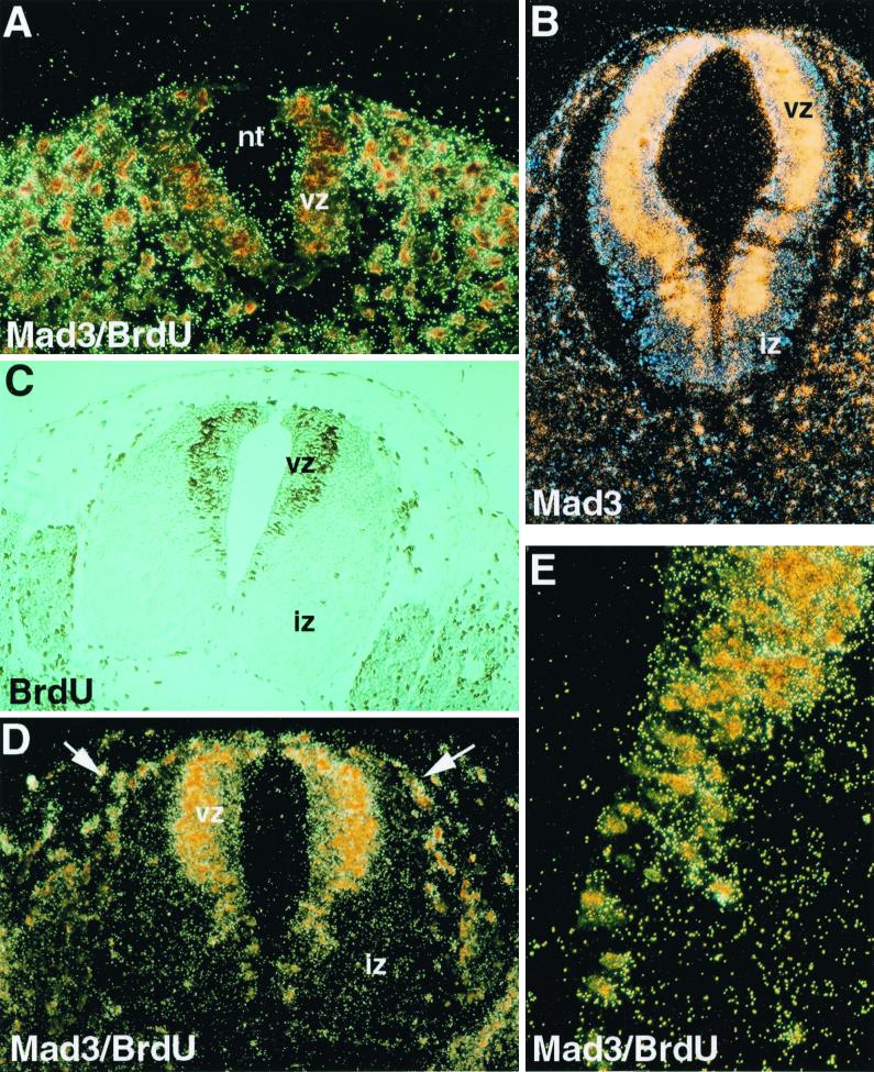
FIG. 1.
Expression of mad3 in neural progenitor cells. (A) Caudal section of a day 11.5 p.c. mouse embryo. The neural tube (nt) shows little sign of differentiation, as evidenced by the absence of the IZ, and consists primarily of proliferating neural progenitors. Brown-stained nuclei indicate cells that have incorporated BrdU for 1 h and are in the S phase of the cell cycle. The section was also hybridized with a mad3 antisense riboprobe. mad3 signal is realtively weakly detected in the proliferating neural progenitors but can be seen in tissue surrounding the neural tube (see panel D). (B) The expression of mad3 is restricted to the periphery of the VZ. No signal is detected in the IZ with the mad3 probe on this day 10.5 p.c. transverse section. (C) Transverse section though the neural tube in a truncal location at day 11.5 p.c. Cells in S phase of the cell cycle are labeled with BrdU (1 h) (brown stain). In the neural tube, the labeled nuclei are localized at the periphery of the VZ. The IZ contains the postmitotic neurons unlabeled with BrdU. Occasional endothelial cells are BrdU labeled. (D) Colocalization of BrdU-positive cells and mad3 in situ hybridization signal. Shown is a transverse section of the neural tube at day 11.5 p.c. processed for immunocytochemistry for the detection of BrdU-incorporating cells and for in situ hybridization to detect mad3. The nuclei at the periphery of the VZ are labeled by BrdU and are also highlighted by the mad3 hybridization signal. Doubly labeled cells can also be observed in the dorsolateral part of the neural tube and are likely to be neural crest cells (arrows). (E) Higher magnification of the VZ pictured in panel D, showing the juxtaposition of mad3 expression and the BrdU-labeled neural progenitors.

