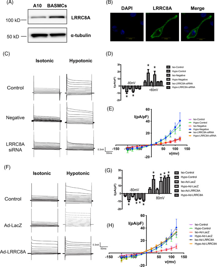
FIGURE 1.

LRRC8A is necessary for VRAC. (A) Representative western blot images showing the expression of LRRC8A in A10 cells and BASMCs. (B) Representative confocal images showing the localization of LRRC8A in BASMCs. Scale, 200 μm. (C–E) Representative traces of VRAC current (C), average current densities measured at ±80 mV (D), and I–V curves (E) in Control, siRNA negative control (Negative), or LRRC8A siRNA‐transfected BASMCs (n = 6, *p < 0.05 VS Control or Negative). (F–H) Representative traces of VRAC current (F), average current densities measured at ±80 mV (G), and I–V curves (H) in Control, Lacz adenovirus (Ad‐Lacz), or LRRC8A‐expressing adenovirus (Ad‐LRRC8A)‐transfected BASMCs (n = 6, *p < 0.05 VS Control or Ad‐LacZ)
