FIGURE 5.

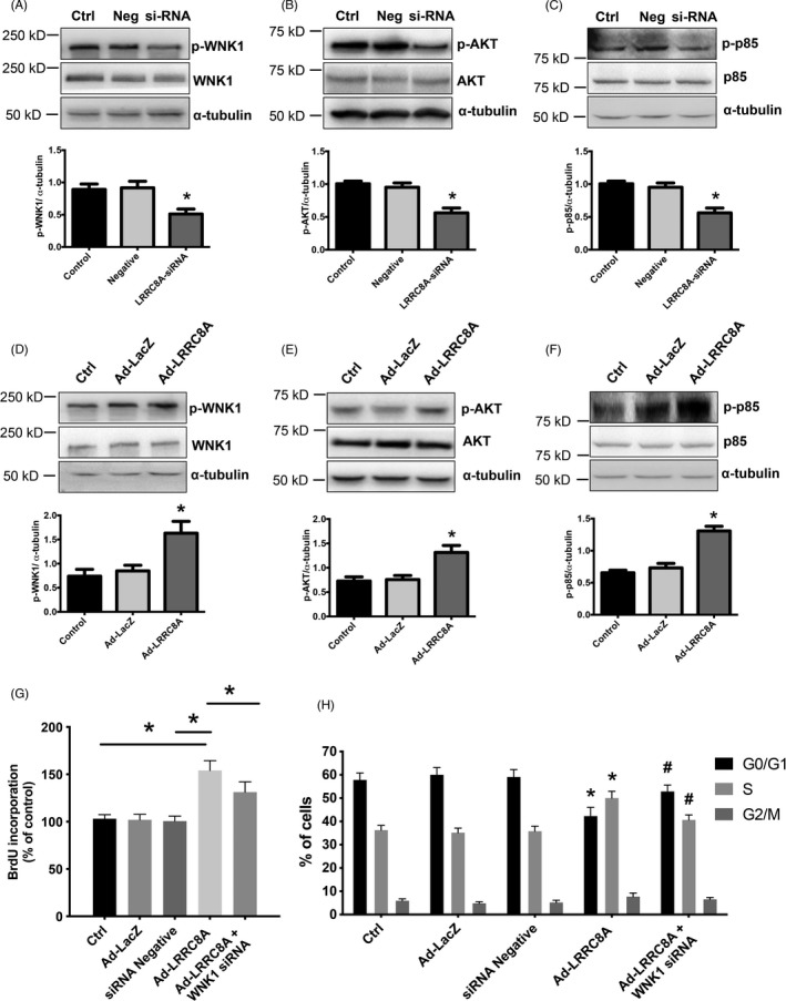
LRRC8A promotes BASMCs proliferation through WNK1/PI3K‐p85/AKT signaling axis. (A–C) Representative western blot images and the respective quantification graphs showing the expression of p‐WNK1 (A), p‐AKT (B), and p‐p85 (C) in LRRC8A siRNA‐treated BASMCs. Ctrl, control; Neg, negative control; si‐RNA, LRRC8A siRNA (n = 5, *p < 0.05 VS Ctrl or Neg). (D–F) Representative western blot images and the respective quantification graphs showing the expression of p‐WNK1 (D), p‐AKT (E), and p‐p85 (F) in LRRC8A siRNA‐treated BASMCs. Ctrl, control; Ad‐LacZ, LacZ control adenovirus; Ad‐LRRC8A, LRRC8A‐expressing adenovirus (n = 6, *p < 0.05 VS Ctrl or Ad‐LacZ). (G) BrdU incorporation assay revealed that overexpression of LRRC8A increased BASMCs proliferation, knock down of WNK1 inhibited the increased effects (n = 5, *p < 0.05). (H) Cell cycle transition was detected by flow cytometric analysis with indicated treatment (n = 4, *p < 0.05 VS Ctrl, # p < 0.05 VS Ad‐LRRC8A)
