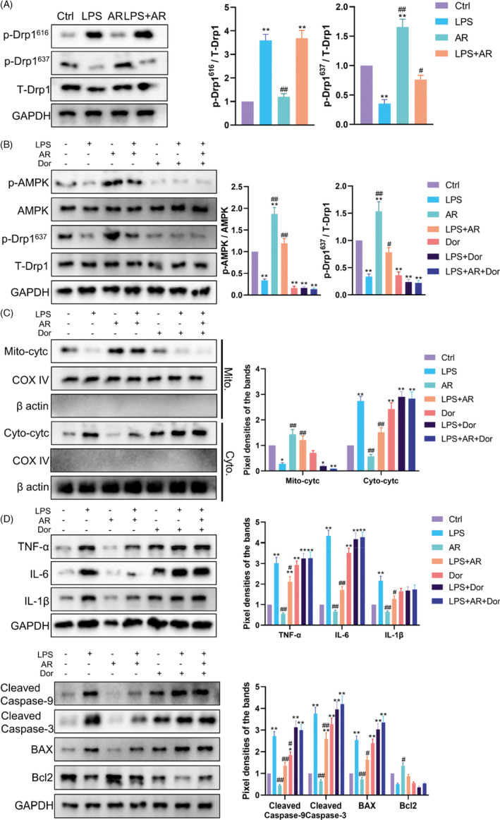
FIGURE 5.

AdipoRon Inhibited Drp1‐mediated Mitochondrial Excessive Division by Phosphorylating AMPK. (A) Western blot was used to measure the phosphorylation level of Drp1 at serine 616 and 637 in ESCs with different treatments. N = 3. * p < 0.05 and ** p < 0.01 versus Ctrl, # p < 0.05 and ## p < 0.01 versus LPS. (B) Western blot was used to measure the phosphorylation level of AMPKα and Drp1 in ESCs with different treatments. N = 3. * p < 0.05 and ** p < 0.01 versus Ctrl, # p < 0.05 and ## p < 0.01 versus LPS. (C) Western blot was used to measure the distribution of Cytochrome C in mitochondria and cytoplasm in ESCs with different treatments. N = 3. * p < 0.05 and ** p < 0.01 versus Ctrl, # p < 0.05 and ## p < 0.01 versus LPS. (D) Western blot was used to measure the inflammation and apoptosis levels in ESCs with different treatments. N = 3. * p < 0.05 and ** p < 0.01 versus Ctrl, # p < 0.05 and ## p < 0.01 versus LPS
