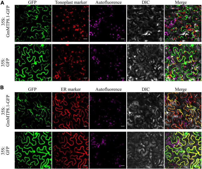
FIGURE 6.
Subcellular localization of GmMTP8.1 in tobacco leaf lower epidermal cells. The images are labeled to show the 35S:GFP and 35S:GFP-GmMTP8.1 constructs. 35S:GFP-GmMTP8.1 was co-expressed with the (A) tonoplast marker TPK1 and (B) endoplasmic reticulum marker PIN5 fused with red fluorescence protein (mKATE) in tobacco leaf epidermal cells. green fluorescent protein (GFP) fluorescence, red fluorescent protein (RFP) fluorescence, chloroplast autofluorescence, bright field images, and merged images are displayed from left to right. Fluorescence was observed by confocal microscopy. Scale bar is 20 μm.

