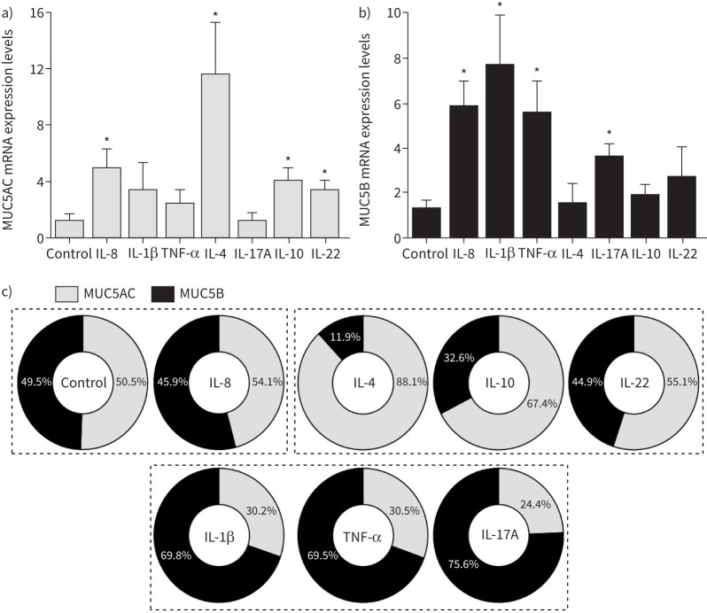
FIGURE 6.
MUC5AC and MUC5B mRNA expression in differentiated BCi-NS1.1 cells following exposure to cytokines. a) MUC5AC and b) MUC5B mRNA expression levels following exposure to cytokines, measured by quantitative reverse transcriptase PCR and normalised to the housekeeping gene GAPDH. Fold-change values are presented as mean±sem, relative to control untreated cells (n=3–5). *: p<0.05 versus control, unpaired t-test. c) Contribution of MUC5AC and MUC5B for mucin expression. Values were calculated by dividing each mucin's fold-change expression levels by the sum of the MUC5AC and MUC5B expression levels shown in a) and b). Dashed lines indicate groups of treatments that have matching contributions of MUC5AC or MUC5B expression. IL: interleukin; TNF: tumour necrosis factor.

