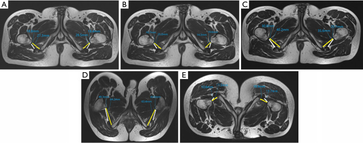
Figure 4.
A healthy 29-year-old male volunteer without hip discomfort: (A) adduction, (B) adduction with 30° external rotation, (C) 30° internal rotation, (D) supine with 30° flexion, (E) prone with 30° backward extension. As demonstrated in the figure, the ischiofemoral space (yellow line) and quadratus femoris space (white line) in the backward extension position were at the minimum, but the space between the ischial tuberosity and the femoral tuberosity still existed and there was no abnormality in the quadratus femoris.

