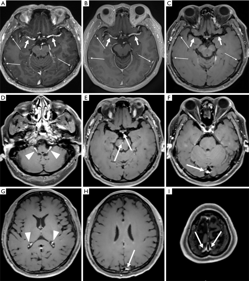
Figure 2.
Representative images obtained using MPRAGE (A), PETRA (B), and DANTE-SPACE (C) in a normal brain, and representative images of DANTE-SPACE at different levels from skull base to head vertex (D,E,F,G,H,I). (A,B) Arterial vessels (short arrows) and small vessels located in brain surface (long arrows) showed signal hyperintensity; (C) the vessels were completely suppressed (short arrows). (E,F,H,I) suppression of some venous vessels was incomplete (long arrows); (D,G) suppression of contrast-enhanced choroid plexus was incomplete (arrowheads). MPRAGE, magnetization-prepared rapid acquisition with gradient echo; PETRA, pointwise encoding time reduction with radial acquisition; DANTE-SPACE, delay alternating with nutation for tailored excitation sampling perfection with application-optimized contrasts using different flip angle evolution.

