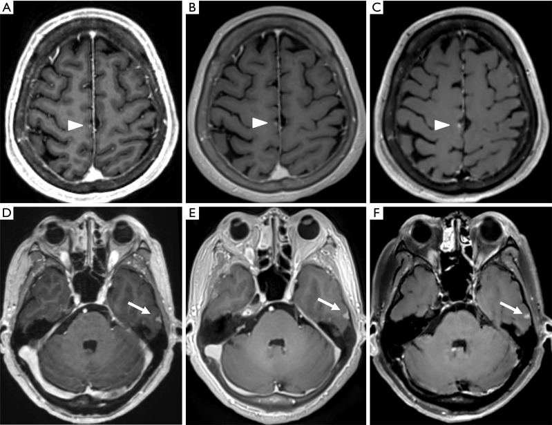
Figure 3.
Representative images of MPRAGE (A,D), PETRA (B,E), and DANTE-SPACE (C,F) of a 57-year-old male patient with cerebral metastases of small-cell lung cancer. In the axial images (A,B,C), the contrast between WM and GM decreased from A to C. A well-enhanced lesion near the cerebral falx in the right frontal lobe can be seen using all three sequences (arrowheads); the lesion appeared to be more visible and slightly larger with DANTE-SPACE than in the MPRAGE and PETRA images. This patient also had a small meningioma in the left temporal lobe (D,E,F); the homogeneously enhanced lesion was displayed clearly with well-defined margins using all three sequences (arrows). MPRAGE, magnetization-prepared rapid acquisition with gradient echo; PETRA, pointwise encoding time reduction with radial acquisition; DANTE-SPACE, delay alternating with nutation for tailored excitation sampling perfection with application-optimized contrasts using different flip angle evolution; WM, white matter; GM, gray matter.

