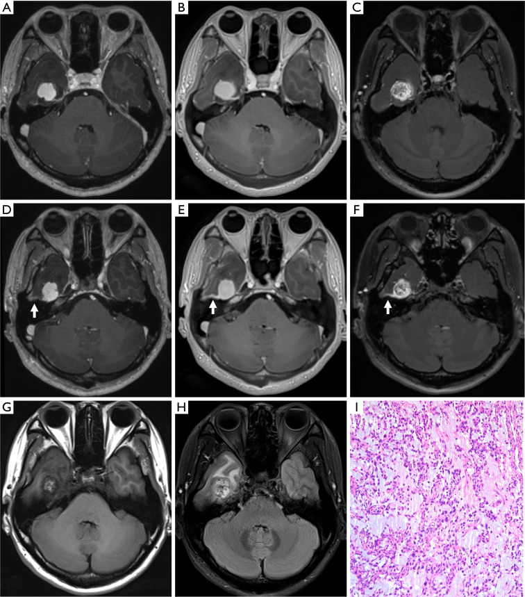
Figure 4.
Post-contrast MPRAGE (A,D), PETRA (B,E), and DANTE-SPACE (C,F) images and pre-contrast T1WI and T2WI (G,H) images of a myopericytoma in the right temporal lobe of a 37-year-old female patient. The signal intensity within this tumor was relatively homogeneous in MPRAGE (A,D) and PETRA (B,E), but was heterogeneous with DANTE-SPACE (C,F). The enhancing effect of adjacent meninges (arrows) was more continuous and more obvious in PETRA (E) compared with MPRAGE (D) and DANTE-SPACE (F). The internal tumor imaging seen in DANTE-SPACE (C,F) was in line with the heterogeneous imaging of pre-contrast images (G,H); this tumor was identified as a myopericytoma by surgical pathology (I, hematoxylin-eosin stain, ×100). MPRAGE, magnetization-prepared rapid acquisition with gradient echo; PETRA, pointwise encoding time reduction with radial acquisition; DANTE-SPACE, delay alternating with nutation for tailored excitation sampling perfection with application-optimized contrasts using different flip angle evolution; T1WI, T1-weighted imaging; T2WI, T2-weighted imaging.

