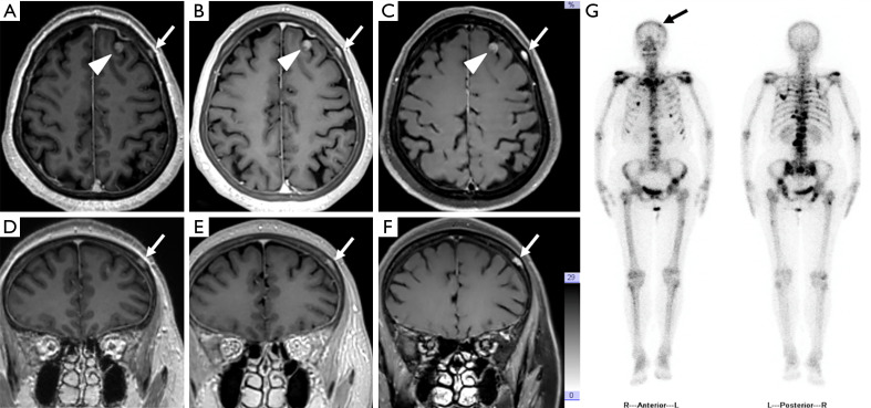
Figure 5.
Post-contrast axial (A,B,C) and coronal (D,E,F) T1WI of a 71-year-old female patient with identified breast cancer obtained using MPRAGE (A,D), PETRA (B,E), and DANTE-SPACE (C,F). The enhancing lesion in the intracranial parenchyma [arrowheads in (A,B,C)] and skull [white arrows in (A,B,C,D,E,F)] were visualized using all three sequences. The detailed structures of the skull diploe were depicted more clearly by PETRA than by MPRAGE and DANTE-SPACE. Anterior and posterior images of whole-body scan by SPECT (G) showed hyperactive metabolism in the spine, ribs, and left frontal-parietal skull (black arrows), suggesting multiple bone metastases. T1WI, T1-weighted imaging; MPRAGE, magnetization-prepared rapid acquisition with gradient echo; PETRA, pointwise encoding time reduction with radial acquisition; DANTE-SPACE, delay alternating with nutation for tailored excitation sampling perfection with application-optimized contrasts using different flip angle evolution; SPECT, single-photon emission computed tomography.

