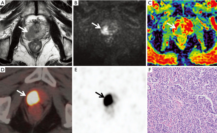
Figure 2.
A 72-year-old patient with increased prostate-specific antigen (PSA) on physical examination (PI-RADS =5, SUVmax =62.4, Gleason score 4+4 =8, PSA: 31 ng/mL). The signal in the right lobe of the prostate decreased unevenly on T2-weighted imaging (T2WI) (A, arrow), a higher signal was observed on diffusion-weighted imaging (DWI) (B, arrow), and a lower signal was noted on the apparent diffusion coefficient (ADC) map (C, arrow). Obvious radioactive uptake was depicted on positron emission tomography/computed tomography (PET/CT) (arrows in D and E). Prostate cancer was proven by pathology (haematoxylin and eosin staining, ×100; F).

