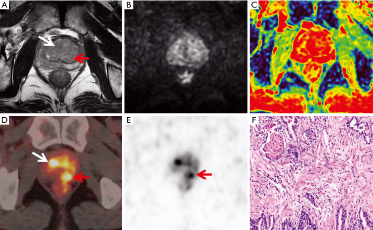
Figure 4.
A 67-year-old patient with increased prostate-specific antigen (PSA) (PI-RADS =2, SUVmax =12.73, PSA: 5.36 ng/mL). A slightly lower signal with blurred borders was visible in the central gland and right peripheral zone on T2-weighted imaging (T2WI) (A, arrows). The diffusion-weighted imaging (DWI) and apparent diffusion coefficient (ADC) maps did not show abnormal signals (B and C). Positron emission tomography/computed tomography (PET/CT) images revealed obvious uptake of diffuse uneven radioactivity (D and E, arrows). Benign prostatic hyperplasia with prostatitis was confirmed by pathology (haematoxylin and eosin staining, 100× magnification; F).

