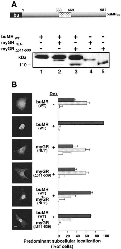
FIG. 9.
MR promotes the nuclear uptake of GR irrespective of GR amino acids 511 to 539. (A). Western blot of COS7 cell extracts comparing the levels of the MR and GR constructs in panel B, using the antibody BuGR. A schematic of the buMR construct is shown at the top of the panel. The GR constructs used are summarized in Fig. 7. (B) In situ immunofluorescence analysis of GR and MR peptide localization in transfected COS7 cells prior to Dex treatment (− Dex) or following 1 h treatment with 10−6 M Dex (− Dex). The localization of the GR constructs prior to hormone treatment is shown in Fig. 7 and is not repeated here. Antibody buGR was used to identify the localization of buMR, while myc epitope antibody 9E10 was used to localize the myGR derivatives in both the absence and presence of buMR. Photomicrographs of the immunofluorescence pattern of representative cells are shown to the left, while quantification of our observations, performed as described in the legend to Fig. 7 is displayed to the right. MR-GR localization in each cell was categorized as completely or mostly nuclear (solid grey bars), equally distributed throughout the cell (stippled bars), or localized predominantly or exclusively to the cytoplasm (white bars). Bar, 10 μm.

