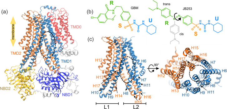
Figure 1.
Model of SUR1 used in simulations (the binding site of glibenclamide (GBM) is shown as a black dashed oval, parts that are unresolved in the original PDB structure are shown in gray, and NBD1 and NBD2 denote the nucleotide binding domains, whereas TMD0, TMD1, and TMD2 stand for the SUR transmembrane domains) (a); structures of the ligands used in this study: GBM and JB253 in cis and trans conformations (b) (blue, orange, and green colors represent the U, S, and R functional parts of ligands, respectively); the transmembrane part of the SUR1 ABC domain in side and top views (c).

