Figure 1.
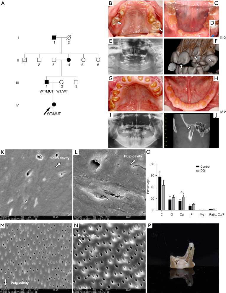
Pedigrees of the DGI family, clinical examinations, and tooth structure investigations. (A) Pedigrees of the DGI family. (B-F) The clinical and radiographic findings for IV.1. (B) Facial and intraoral examinations showing the enamel defect on newly erupted teeth (white arrows). (C,D) Soon after the eruption of the mandibular right first molar, a fistula appeared (blue arrow). (E) The oral panoramic radiographs show that the teeth pulp cavities of unerupted teeth were available, while the cavities of the erupted teeth were partially obliterated. (F) The CBCT image of the unerupted teeth shows hypoplastic enamel defects (white arrows). (G-J) The clinical and radiographic finding for of III.1. (G,H) Teeth are yellow-brownish colored and show severe attrition to the gingiva margin. (I) The oral panoramic radiographs show that the teeth pulp cavities are totally obliterated, and the roots are short and round. (J) The CBCT image shows that the 2 mandibular heads are asymmetric; the left mandibular head shows attrition and is rotated forward (white arrow). (K) SEM image of the DGI tooth (2,000×). (L) SEM image of the DGI tooth (5,000×). (M) SEM image of the control (2,000×). (N) SEM image of the control (5,000×). (White arrows indicate the direction of pulp cavity). (O) EDS results of the DGI tooth and the control, *, P<0.05. (P) Image of the DGI tooth. DGI, dentinogenesis imperfecta; CBCT, cone-beam computed tomography; SEM, scanning electron microscopy; EDS, energy dispersive spectroscopy.

