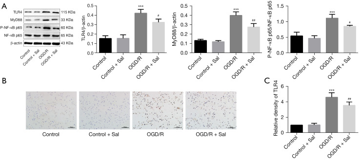
Figure 5.
Sal inhibited the TLR4/NF-κB signaling pathway activation in OGD/R-induced BV2 cells. (A) Protein expression levels of TLR4, MyD88, p-NF-κB p65, and NF-κB p65 were detected by western blot (n=3); representative bands are shown in the figure. (B) Protein expression levels of TLR4 were detected by IHC staining (scale bar =100 µm). (C) Quantitative analysis of IHC staining results (n=5). Data are expressed as mean ± SD. ***P<0.001 compared with control group; #P<0.05, ##P<0.01 compared with OGD/R group. OGD, oxygen and glucose deprivation; TLR4, Toll-like receptor 4; MyD88, myeloid differentiation primary response 88; p-NF-κB p65, phosphorylated nuclear factor kappa B p65; IHC, immunohistochemical; SD, standard deviation.

