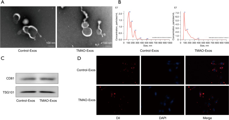
Figure 1.
Isolation and characterization of Exos isolated from hepatocytes culture supernatant. (A) Nanovesicles with diameters of around 100 nm were isolated and purified from the hepatocyte culture supernatant, and visualized under an electron microscope. (B) The size distribution of these nanovesicles were further identified by nanoparticle tracking analysis. (C) The expressions of exosomal identity marker CD81 and TSG101 were detected in the Control-Exos and TMAO-Exos by western blotting. (D) The Exos were labelled with DiI and co-cultured with HAECs for 24 h. The results showed that the DiI-labelled Exos could be taken up by HAECs (400× magnification). Exos, exosomes; CD81, cluster of differentiation 81; TSG101, tumor susceptibility gene 101; TMAO, trimethylamine-N-oxide; HAECs, human aortic endothelial cells.

