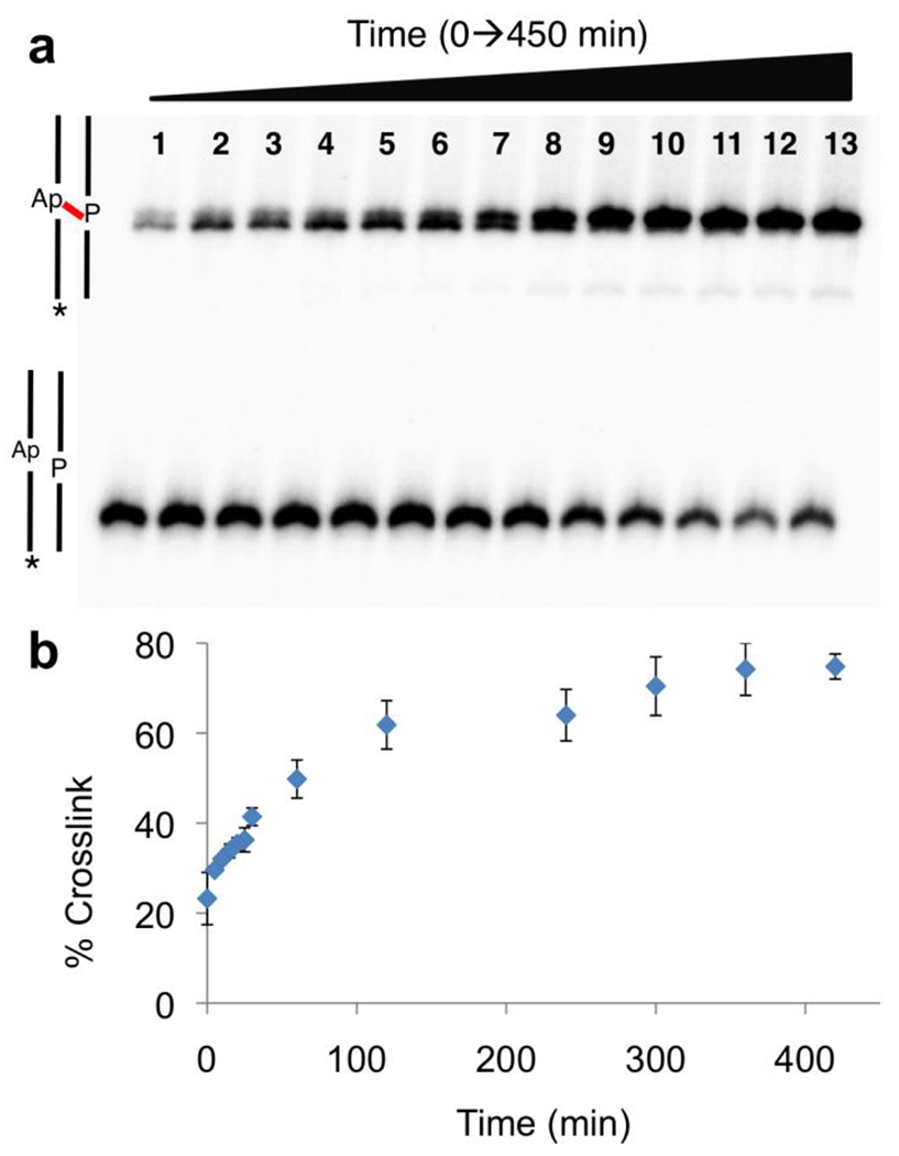
Figure 2. Time-course for the formation of the P-Apred in duplex B.

Panel a: The Ap-containing duplex was incubated in sodium acetate buffer (750 mM, pH 5.2) containing NaCNBH3 (250 mM) at 37 °C. Aliquots were removed at 0, 5, 10, 15, 20, 25, 30, 60, 120, 240, 300, 360 and 420 min and frozen prior to gel analysis. The 32P-labeled 2’-deoxyoligonucleotides were resolved on a 20% denaturing polyacrylamide gel and the radioactivity in each band quantitatively measured by storage-phosphor autoradiography. The fast-migrating band corresponds to the 32P-labeled full-length Ap-containing strand from uncross-linked duplex and the upper, slowly-migrating band corresponds to the cross-linked DNA duplexes. Panel b: Plot of cross-link yield versus time derived from the gel electrophoretic data (shown in Panel a).
