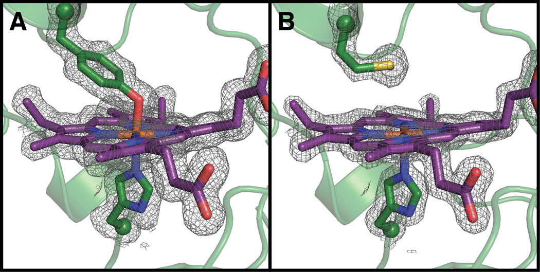
Figure 2.
Compared to the wild-type structure (A; PDB ID: 6NX0), the Y463M mutant (B, PDB ID: 6V59) disrupts axial heme ligation. 2FO – FC composite omit electron density countoured at 1 σ Peptide and heme carbon shown in green and purple, respectively. Oxygen, nitrogen, sulfur, and iron shown in red, blue, yellow, and orange, respectively. Peptide backbone shown in ribbons representation and side chains and hemes shown in stick representation. Side chain α-carbon shown as spheres. Terminal methyl group of Met side chain is not visible in this orientation. 2FO – FC electron density shown in grey and contoured at 1σ.

