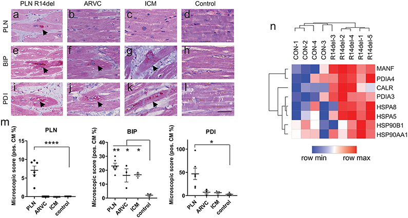
Figure 3. Determination of the UPR status in PLN R14del disease and other forms of cardiomyopathies.
Histological analysis of human myocardium from patients suffering from PLN R14del, desmosomal arrhythmogenic cardiomyopathy (ARVC), and ischemic cardiomyopathy (ICM) versus control (healthy) hearts.
a–d, Abnormal accumulation of PLN in perinuclear aggregates (arrows) in severely affected cardiomyocytes in PLN R14del and absent in ARVC, ICM, control.
e-h, Diffuse moderate immunolabeling (arrows) for BIP in PLN 14del, and present to a lesser extent in ARVC and ICM.
i-l, High immunolabeling for dotted cytoplasmic PDI (arrows) in PLN 14del, low PDI presence in ARVC, and ICM (scale bar = 25 μm).
m, Quantification of immunostaining in PLN R14del (n = 6 patients), ARVC (n = 3 patients), ICM (n = 3 patients), and control (n = 3 patients).
n, RNA sequence analysis of UPR gene expression from healthy and PLN R14del myocardium from patient samples.
Data were presented as mean ± SEM. * p < 0.05, ** p < 0.005, **** p < 0.00005.

